Urorectal septum
Urorectal septum
Jump to navigation
Jump to search
| Urorectal septum | |
|---|---|
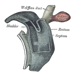
 Cloaca of human embryo from twenty-five to twenty-seven days old. | |
| Details | |
| Days | 32 |
| Identifiers | |
| Latin | septum urorectale |
| TE | E5.4.9.0.2.0.14 |
Anatomical terminology [edit on Wikidata] | |
The endodermal cloaca is divided into a dorsal and a ventral part by means of a partition, the urorectal septum, which grows downward from the ridge separating the allantois from the cloacal opening of the intestine and ultimately fuses with the cloacal membrane and divides it into an anal and a urogenital part. The dorsal part of the cloaca forms the rectum, and the anterior part of the urogenital sinus and bladder. The urorectal septum eventually becomes part of the perineal body.
Clinical significance[edit]
Malformation of the urorectal septum can lead to several different types of fistulas.[1]
References[edit]
This article incorporates text in the public domain from page 1109 of the 20th edition of Gray's Anatomy (1918)
^ Escobar LF, Heiman M, Zimmer D, Careskey H (2007). "Urorectal septum malformation sequence: Prenatal progression, clinical report, and embryology review". Am. J. Med. Genet. A. 143 (22): 2722–6. doi:10.1002/ajmg.a.31925. PMID 17937427..mw-parser-output cite.citationfont-style:inherit.mw-parser-output .citation qquotes:"""""""'""'".mw-parser-output .citation .cs1-lock-free abackground:url("//upload.wikimedia.org/wikipedia/commons/thumb/6/65/Lock-green.svg/9px-Lock-green.svg.png")no-repeat;background-position:right .1em center.mw-parser-output .citation .cs1-lock-limited a,.mw-parser-output .citation .cs1-lock-registration abackground:url("//upload.wikimedia.org/wikipedia/commons/thumb/d/d6/Lock-gray-alt-2.svg/9px-Lock-gray-alt-2.svg.png")no-repeat;background-position:right .1em center.mw-parser-output .citation .cs1-lock-subscription abackground:url("//upload.wikimedia.org/wikipedia/commons/thumb/a/aa/Lock-red-alt-2.svg/9px-Lock-red-alt-2.svg.png")no-repeat;background-position:right .1em center.mw-parser-output .cs1-subscription,.mw-parser-output .cs1-registrationcolor:#555.mw-parser-output .cs1-subscription span,.mw-parser-output .cs1-registration spanborder-bottom:1px dotted;cursor:help.mw-parser-output .cs1-ws-icon abackground:url("//upload.wikimedia.org/wikipedia/commons/thumb/4/4c/Wikisource-logo.svg/12px-Wikisource-logo.svg.png")no-repeat;background-position:right .1em center.mw-parser-output code.cs1-codecolor:inherit;background:inherit;border:inherit;padding:inherit.mw-parser-output .cs1-hidden-errordisplay:none;font-size:100%.mw-parser-output .cs1-visible-errorfont-size:100%.mw-parser-output .cs1-maintdisplay:none;color:#33aa33;margin-left:0.3em.mw-parser-output .cs1-subscription,.mw-parser-output .cs1-registration,.mw-parser-output .cs1-formatfont-size:95%.mw-parser-output .cs1-kern-left,.mw-parser-output .cs1-kern-wl-leftpadding-left:0.2em.mw-parser-output .cs1-kern-right,.mw-parser-output .cs1-kern-wl-rightpadding-right:0.2em
External links[edit]
- http://cancerweb.ncl.ac.uk/cgi-bin/omd?urorectal+septum
- http://www.embryology.ch/anglais/turinary/urinbasse01.html
- http://isc.temple.edu/marino/embryology/parch98/parch_text.htm
- http://www.med.mun.ca/anatomyts/renal/akid5.htm
- http://www.med.umich.edu/lrc/coursepages/M1/embryology/embryo/10digestivesystem.htm
This developmental biology article is a stub. You can help Wikipedia by expanding it. |
Categories:
- Wikipedia articles incorporating text from the 20th edition of Gray's Anatomy (1918)
- Embryology of digestive system
- Developmental biology stubs
(window.RLQ=window.RLQ||).push(function()mw.config.set("wgPageParseReport":"limitreport":"cputime":"0.256","walltime":"0.373","ppvisitednodes":"value":1439,"limit":1000000,"ppgeneratednodes":"value":0,"limit":1500000,"postexpandincludesize":"value":21637,"limit":2097152,"templateargumentsize":"value":919,"limit":2097152,"expansiondepth":"value":25,"limit":40,"expensivefunctioncount":"value":4,"limit":500,"unstrip-depth":"value":1,"limit":20,"unstrip-size":"value":3371,"limit":5000000,"entityaccesscount":"value":3,"limit":400,"timingprofile":["100.00% 328.063 1 -total"," 41.28% 135.422 1 Template:Infobox_embryology"," 39.86% 130.751 1 Template:Infobox_anatomy"," 36.25% 118.917 1 Template:Infobox"," 27.58% 90.494 1 Template:Reflist"," 25.64% 84.104 1 Template:Cite_journal"," 11.93% 39.128 1 Template:TerminologiaEmbryologica"," 10.19% 33.443 1 Template:Gray's"," 10.02% 32.882 3 Template:Str_mid"," 9.55% 31.317 2 Template:Wikidata"],"scribunto":"limitreport-timeusage":"value":"0.128","limit":"10.000","limitreport-memusage":"value":3801975,"limit":52428800,"cachereport":"origin":"mw1332","timestamp":"20190420160335","ttl":2592000,"transientcontent":false););"@context":"https://schema.org","@type":"Article","name":"Urorectal septum","url":"https://en.wikipedia.org/wiki/Urorectal_septum","sameAs":"http://www.wikidata.org/entity/Q7901043","mainEntity":"http://www.wikidata.org/entity/Q7901043","author":"@type":"Organization","name":"Contributors to Wikimedia projects","publisher":"@type":"Organization","name":"Wikimedia Foundation, Inc.","logo":"@type":"ImageObject","url":"https://www.wikimedia.org/static/images/wmf-hor-googpub.png","datePublished":"2007-11-07T04:03:00Z","dateModified":"2018-11-27T19:59:42Z","image":"https://upload.wikimedia.org/wikipedia/commons/f/fb/Gray992.png"(window.RLQ=window.RLQ||).push(function()mw.config.set("wgBackendResponseTime":106,"wgHostname":"mw1270"););

