Parotid duct
| Parotid duct | |
|---|---|
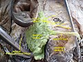
 Right parotid gland. Deep and anterior aspects. (Parotid duct labeled at center left.) | |
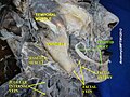
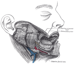 Dissection, showing salivary glands of right side. (Parotid duct visible at center.) | |
| Details | |
| Identifiers | |
| Latin | Ductus parotideus |
| MeSH | D018987 |
| TA | A05.1.02.007 |
| FMA | 10420 |
Anatomical terminology [edit on Wikidata] | |
The parotid duct or Stensen duct is a duct and the route that saliva takes from the major salivary gland, the parotid gland into the mouth.[1]
Contents
1 Eponym
2 Structure
3 Clinical relevance
4 Additional images
5 See also
6 References
7 External links
Eponym
It is named after Nicolas Steno (1638–1686), a Danish anatomist credited with its detailed description in 1660.
Structure
The parotid duct is formed when several interlobular ducts—the largest ducts inside the parotid gland—join. It emerges from the gland and runs forward along the lateral side of the masseter muscle. In this course, the duct is surrounded by the buccal fat pad.[2] It takes a steep turn at the border of the masseter and passes through the buccinator muscle, opening into the vestibule of the mouth, the region of the mouth between the cheek and the gums, at the parotid papilla, which lies across the second superior molar tooth.[3]
The buccinator acts as a valve that prevents air forcing into the duct, which would cause pneumoparotitis.[4] Running along with the duct superiorly is the transverse facial artery and upper buccal nerve; running along with the duct inferiorly is the lower buccal nerve.
The exit of the parotid ducts can be felt as small bumps (Papillae) on both sides of the mouth, and are usually positioned next to the maxillary second molars.
Clinical relevance
Blockage, whether caused by salivary duct stones or external compression, may cause pain and swelling of the parotid gland (parotitis).
Koplik's spots which are pathognomonic of measles are found near the opening of the parotid duct.
Additional images
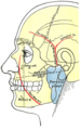
Outline of side of face, showing chief surface markings.
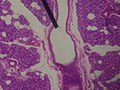
Microscopic slide of a human interlobular duct.
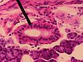
Microscopic slide of a human striated duct.

The left papilla (soft tissue protuberance at the exit) of the parotid duct is clearly visible on the cheek in the right of the photo.
Parotid duct
Parotid duct

Parotid duct


See also
- Parotid gland
- Parotitis
References
^ Ten Cate's Oral Histology, Nanci, Elsevier, 2013, page 255
^ Latarjet, Michel; Ruiz Liard, Alfredo (2005). Human Anatomy (Spanish Edition). Editorial Médica Panamericana. ISBN 978-950-06-1368-2..mw-parser-output cite.citationfont-style:inherit.mw-parser-output qquotes:"""""""'""'".mw-parser-output code.cs1-codecolor:inherit;background:inherit;border:inherit;padding:inherit.mw-parser-output .cs1-lock-free abackground:url("//upload.wikimedia.org/wikipedia/commons/thumb/6/65/Lock-green.svg/9px-Lock-green.svg.png")no-repeat;background-position:right .1em center.mw-parser-output .cs1-lock-limited a,.mw-parser-output .cs1-lock-registration abackground:url("//upload.wikimedia.org/wikipedia/commons/thumb/d/d6/Lock-gray-alt-2.svg/9px-Lock-gray-alt-2.svg.png")no-repeat;background-position:right .1em center.mw-parser-output .cs1-lock-subscription abackground:url("//upload.wikimedia.org/wikipedia/commons/thumb/a/aa/Lock-red-alt-2.svg/9px-Lock-red-alt-2.svg.png")no-repeat;background-position:right .1em center.mw-parser-output .cs1-subscription,.mw-parser-output .cs1-registrationcolor:#555.mw-parser-output .cs1-subscription span,.mw-parser-output .cs1-registration spanborder-bottom:1px dotted;cursor:help.mw-parser-output .cs1-hidden-errordisplay:none;font-size:100%.mw-parser-output .cs1-visible-errorfont-size:100%.mw-parser-output .cs1-subscription,.mw-parser-output .cs1-registration,.mw-parser-output .cs1-formatfont-size:95%.mw-parser-output .cs1-kern-left,.mw-parser-output .cs1-kern-wl-leftpadding-left:0.2em.mw-parser-output .cs1-kern-right,.mw-parser-output .cs1-kern-wl-rightpadding-right:0.2em
^ Illustrated Dental Embryology, Histology, and Anatomy, Bath-Balogh and Fehrenbach, Elsevier, 2011, page 135
^ Faizal B, Chandran MP (2012). "Pneumoparotitis" (PDF). Amrita Journal of Medicine. 8 (2): 1–44. Archived from the original (PDF) on 2015-12-11.
Casseri, Giulio Cesare (1627). Tabulae anatomicae, lxxiix. Venetis.
Stensen, Niels (1662). Observationes anatomicae, quibus varia oris, oculorum & narium vas describuntur novique salivae, lacrymarum & muci fontes deteguntur. Lugduni Batavorum: J. Chouët.
External links
- Diagram at MSU
ent/178 at eMedicine - Parotid duct injuries
doctor/2052 at Who Named It?
Authority control |
|
|---|

